


Radiology Department
แผนกรังสีวิทยา
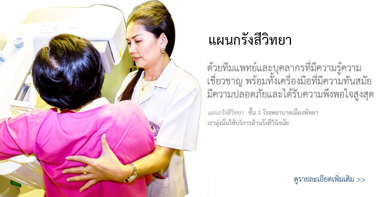
Diagnostic Center X-ray, Ultrasound, mammogram
Welcome to Pattaya City Hospitals Diagnostic Center
Ultrasound Machine
When an ultrasound scan may be used
An ultrasound scan can be used in several different ways, such as monitoring an unborn baby, diagnosing a condition or guiding a surgeon during certain procedures.
Pregnancy
Ultrasound scans are a routine procedure for pregnant women. They produce images of the unborn baby inside the womb and display them on a monitor.
Most women are offered at least two ultrasound scans during pregnancy:
• the first scan (at around 8-14 weeks) can help to confirm the pregnancy and determine when the baby is due
• the second scan (usually at around 18-21 weeks) checks for structural abnormalities, particularly in the baby's head or spine
However, an ultrasound scan can be done at any time during pregnancy and causes no harm to the baby.
Read more information about when ultrasound is used during pregnancy.
Diagnosing conditions
Ultrasound scans can help diagnose problems in many parts of your body, including your:
- liver (cirrhosis)
- gallbladder (gallstones)
- thyroid gland
- lymph nodes
- ovaries
- uterus (womb)
- testes
- breasts
For example, it can help to detect whether a lump in one of these organs is a tumour or a cyst.
Ultrasound may also be used to diagnose problems with your:
- blood vessels (aneurysm)
- joints, ligaments and tendons
- skin
- eyes
The hip, spine and brain of newborn babies can be scanned for abnormalities, but by 18 months old the skull has fully grown and it is no longer possible to use ultrasound on the brain without surgery.
Mammogram
Women between the ages of 20 and 39 who have no symptoms of breast disease should have a clinical breast examination every 3 years. A physician, a nurse practitioner, a nurse, or a physician assistant can perform this examination. The health care professional will examine both breasts for discrepancies in size or shape. The examiner will also palpate (feel) each breast to detect any lumps or masses. The area under both arms will be examined as well, to check for enlargement of lymph nodes.
Mammography
A mammogram is an x-ray examination of the breasts, used to detect and diagnose breast diseases. Mammography is the most effective method of detecting cancer at an early stage, before the woman or a physician can feel it.
Screening mammography is used as a preventive measure for women who have no symptoms of breast disease. A screening mammogram usually involves two views of each breast. The American Cancer Society recommends that all women aged 40 and over have a screening mammogram every year as part of a breast health program, which also includes an annual breast examination by a healthcare professional.
Diagnostic mammography involves additional views of the breast, and is used when an abnormality is found during screening, or in women who have breast complaints, such as a breast mass, nipple discharge, breast pain, or skin irritation.
Mammography Procedure
When a woman has a mammogram, she must undress above the waist and wear an open wrap, which is provided by the mammography facility. A breast-imaging technologist will place the patient in front of the machine and position each breast, one at a time, on the mammography equipment. The technologist will position the patient and the breast in the appropriate location for obtaining the best image of the breast. A paddle is then used to gently compress the breast so that the tissue is flattened.
Compressing the breast is necessary to obtain the best image. Compression thins and evens the breast tissue, so that a lower x-ray dose can be used. Even distribution of breast tissues reduces x-ray scatter, or the spread of radiation from the targeted tissue, which provides a better quality image. Compression also prevents the breast from moving during the procedure, thus reducing or eliminating blurred images. Breast compression lasts for a few seconds. It is uncomfortable, but should not be painful.
Some women find breast compression to be more uncomfortable than others; in fact, some women avoid having mammograms because they fear it will be painful or they had a past experience that was painful. There is a breast cushion, the MammoPad, that can be used during the exam to reduce the discomfort associated with mammography.
When the breast is positioned and compression is complete, the technologist will leave the room or step behind a screen. Once shielded, the technologist will turn on the x-ray source to transfer the image of the breast onto the x-ray film or the digital detector. The process of positioning and radiating is repeated for additional views of the same breast and for the other breast.
If a screen-film examination is being conducted, the technologist will take all of the x-rays needed for the examination. She will then develop the films before the patient leaves, to make sure each film shows the right view and exposure.
In digital mammograms, the image for each exposure will appear on the technologist's computer screen, and she will be able to make sure each image is acceptable before positioning the patient for the next view.
The entire procedure for screening mammography should take 15 to 20 minutes for a screen-film examination and 5 to 10 minutes for digital mammography. The procedure may take longer if additional views are needed, as is the case in diagnostic mammography or for mammography of women with breast implants. Breast implants create a unique imaging situation, because some breast tissue will be covered by the implant and cannot be seen on x-rays. To compensate for this, additional films are taken, to allow as much breast tissue as possible to be imaged.
The mammography films are interpreted by a radiologist, who will look for abnormalities and compare the new films to previous mammograms, to detect any changes. The radiologist prepares a report for your mammography facility. The mammography facility is responsible for notifying the patient of the results.
ตารางแพทย์ออกตรวจ:

<< Back to menu
Copyright © 2015 PATTAYA CITY HOSPITAL All RIGHTS RESERVED
261/40 S. Bua Khaow Nongprue, Banglamung, 20150
CallCenter: 038-103-900 Fax: 038-103-912
E-mail:- cs.pch@hotmail.com
E-mail:- cs.pch@hotmail.com
![]()
![]()